
Home / Procedures & Investigations / Dermoscopy
Dermoscopy
Dermoscopy is a non-invasive skin examination that uses magnification and light to assess moles, lesions, and other skin concerns helping identify early signs of skin cancer and other dermatological conditions.
How does Dermoscopy work?
Dermoscopy (also known as dermatoscopy or skin surface microscopy) is examination technique used to assess skin lesions or moles more closely.
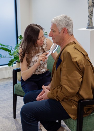
A handheld device called a dermatoscope provides magnification and light, helping your specialist see structures in the skin not visible to the naked eye.
A dermoscopy is used to help diagnose skin cancers, including melanoma, and to monitor changes in moles over time.
The Examination
The dermatoscope provides high magnification of the skin’s surface, allowing your specialist to examine fine details such as pigmentation, blood vessels, and skin texture.
It uses a powerful light source to illuminate the skin, which helps clinicians see subsurface structures and patterns.
Your specialist will advise you if this test might help guide your personalised treatment regime.
Step 1
When you arrive for the test you may be asked to change into a hospital gown. Your specialist will take care to put you at ease and to ensure you’re as comfortable as possible for the procedure.
Step 2
During the examination, your specialist will place the dermatoscope directly on your skin. The procedure is quick and painless.
Step 3
The specialist may examine one or several skin lesions, and may take photographs for future comparison. If any lesions are suspicious, you may be advised to have a biopsy or follow-up appointment.
Find A Specialist
-

Dr Amanda Peacock
Plastic & Reconstructive Surgeon
